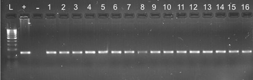
Figure 1: Electrophoresis gel showing 100bp DNA ladder (L), positive control (+) lane, empty negative control lane (–), and samples 1-16 as indicated in the corresponding lanes.
Comparative Gene Approach to the Investigation of SNPs within the Tenascin-C Gene in Achilles Tendon Injury in the Canine Patient
Nina R. Kieves DVM, DACVS-SA, DACVSMR, CCRT1*
1Department of Veterinary Clinical Sciences, The Ohio State University, 601 Vernon L Tharp St, Columbus, OH 43210
2University of Illinois, Veterinary Clinical Medicine, 1008 W Hazelwood Dr, Urbana, IL 61802
3Glenwood City Veterinary Clinic, 308 Syme Ave, Glenwood City, WI 54013
*Corresponding Author (nkieves@gmail.com)
Vol 1, Issue 3 (2016)
Published: 04 August 2016
Reviewed by: Matthew Stewart (MVet, PhD)
DOI: 10.18849/VE.V1I3.36
Objective: To evaluate for single nucleotide polymorphisms (SNPs) within the tenascin-C (TNC) gene in a population of dogs with atraumatic Achilles tendon rupture.
Background: In humans, Achilles tendinopathy has been extensively studied for numerous polymorphisms within several genes and has been associated with polymorphisms in collagen (COL5A1) and the TNC genes.
Evidentiary value: This study serves as a starting point for evaluating a genetic component of Achilles tendinopathy in the dog.
Methods: Whole blood from twenty dogs with atraumatic Achilles tendon rupture and 14 matched control samples were used. DNA was extracted from whole blood run with primers designed around two SNPs previously identified to be related to Achilles tendinopathy in humans. One SNP was located in exon 29, and one exon 17 of the canine TNC gene. Polymerase chain reaction (PCR) was run on the samples and they were sequenced. Sequences of the affected canine population were compared to the control sample sequences.
Results: There were no significant differences in genotype or allele frequency of the SNPs rs13321 and rs2104772 between any of the affected and control subjects with a p-value of 1.0.
Conclusion: This study evaluated a population of canines with atraumatic Achilles tendon rupture for SNPs in the TNC gene. We found no difference in gene sequence for the study population compared to age, sex, and breed matched controls.
Application: Though the data from this study did not show a correlation between the specific polymorphisms investigated, it is possible that other SNPs within the TNC gene or other genes involved in tendon composition and repair such as collagen may be associated with atraumatic Achilles tendon injury in the dog.
The common calcanean tendon, or Achilles tendon, is comprised of three main components; the paired tendon of the gastrocnemius muscles; the combined tendon of the gracilis, semitendinosus, and biceps femoris muscles; and the tendon of the superficial digital flexor muscle. Tendons are exposed to high mechanical stresses, and disruption can be partial or complete. In the canine patient, disruption can be secondary to a traumatic rupture, often involving direct laceration of the tendon, or can be atraumatic or spontaneous with no distinct injury or event noted1,2.
Intrinsic and extrinsic factors can affect an individual’s risk for developing a tendinopathy. Extrinsic factors shown to be associated with Achilles tendon rupture in humans include type of training program, poor technique, type of footwear, previous injuries sustained, and environmental factors3. Intrinsic risk factors for developing tendinopathy, including genetic variations in chromosome 9, are well documented in humans4-6 . In humans, Achilles tendinopathy has been extensively studied for numerous polymorphisms within several genes and has been found to be associated with polymorphisms in collagen (COL5A1) and the tenascin genes4,7-16. Tenascin encodes the extracellular matrix tenascin-C glycoprotein, which is expressed during wound healing and remodeling of adult tissues, especially those exposed to high tensile and compressive stress. A 2012 study by Saunders et al., evaluated a number of SNPs within the human TNC gene of patients affected with Achilles tendon injury7. They found that several SNPs were significantly associated with an increased risk of acute Achilles tendon injury in a South African and Australian study population when compared to asymptomatic controls7.
In the canine, traumatic lacerations of the Achilles tendon are often primarily repaired, followed by a period of immobilization of the hock joint. Atraumatic injuries, however, are often insidious, and can be frustrating to treat successfully often causing decreased function to the patient in the long-term. If there is a genetic mutation predisposing dogs to atraumatic Achilles tendon injuries, this could help target therapies to treat these patients more successfully, and possibly allow screening for at risk patients.
To the author’s knowledge, no investigation has been made into a genetic etiopathogenesis of atraumatic Achilles tendon injury in the canine. Therefore, the objective of our study was to evaluate the canine TNC gene for SNPs rs13321 and rs2104772 previously identified to be expressed in human populations undergoing atraumatic Achilles tendon injury. We hypothesised that there would be expression of a similar SNP in the canine genome in dogs with atraumatic Achilles tendon injury in comparison to an age, sex, and breed matched control group.
Sample Collection
Electronic medical records at the University of Minnesota Veterinary Medical Center were retrospectively reviewed for Achilles tendon injuries between 2005-2010. Dogs with traumatic rupture were excluded. Dogs were included if they had been diagnosed with an atraumatic Achilles tendon injury, and were alive and available to return to the hospital, where whole blood samples were obtained, with owner consent. Age, breed, and gender matched control samples were obtained from a DNA storage bank maintained at the University of Minnesota Surgical Research Laboratory.
DNA Isolation
DNA was extracted from approximately 10 mls of whole blood using the QIAamp DNA Blood Midi/Maxi Kita. Protocol, as determined from the January 2005 handbook22, is to lyse the red blood cells, bind the DNA to a spin column, wash with multiple buffers, and then elute the DNA into nuclease-free water and store at -20°C. Concentration and purity of the DNA were assessed with a spectrophotometerb.
Primer Sequence
Primers were designed using Primer 3 Plusc. Primers were designed around two SNPs identified and investigated by Saunders et al. in 20127. The BLAST algorithm was used to compare the human TNC SNPs rs13321 and rs2104772 sequence determined by National Center for Biotechnology Information SNP databased to the canine genome; the BLAST algorithm revealed the synonymous sequences. The synonymous canine coding region surrounding SNP rs13321 was located in exon 29 of the canine TNC gene and the synonymous canine coding region of SNP rs2104772 was located in exon 17 of the canine TNC genome. The sequences of the exons were inputed into Primer 3 for primer design; the forward and reverse primers were then forced to include the known SNPs (Table 2).
PCR and Gel Electrophoresis
For this reaction, a 10 ml PCR volume was prepared that contained approximately 12.5 ng of genomic canine DNA, 2.5 pmol of each primer, 0.20 mM dNTP, 2.0 ml of 5X GoTaq Reaction Buffer, 1 mM MgCl2, 0.05 U GoTaq Hot Start Polymerase and distilled, nuclease-free water to bring the total volume to 10 ml. A 269 bp the product was amplified using a thermocyclere. The thermal cycling protocol was set to an initial denaturing for 3 minutes at 94°C, 35 cycles at 94°C for 30 seconds denaturing, 61.1°C for 45 seconds annealing, and 72°C for 45 seconds of extension, with a final extension of 10 minutes at 72°C. PCR products were stored at 4°C for less than 24 hours until gel electrophoresis was run. Two ml of each PCR product was run on a 2% agarose gelf for 1 hour at 109v. A 100 bp DNA ladderg with size markers was used to confirm correct product size (Figure 1).
Product Sequencing
Once correct products were confirmed, the PCR products were cleaned with ExoSAP-ITh. ExoSAP-IT reagent (3.2 ml) was added to each PCR product and then placed in the thermal cycler for a program of 15 minutes at 80°C and then 15 minutes at 37°C. In preparation for sequencing, 2 ml of each PCR product was prepared with 9 ml of nuclease-free water and 25 pmol of primer (forward in one preparation, reverse in the other). Samples were sent to Biomedical Genomics Center (BMGC)i to be sequenced using Sanger Sequencing. Results were analysed with Sequencher softwarej.
Statistics
A Fisher’s exact statistical test was performed utilising commercially available softwarek to compare the cases and controls. A p-value of <0.05 was considered statistically significant.
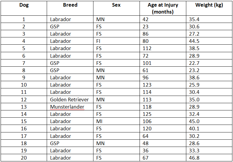
Twenty dogs met the inclusion criteria for the study. The mean age was 7.1 years ± 2.4 (range 2-10 years), and mean weight was 33.1kg ± 7.1 (range 22.7-46.8kg). Breeds affected included Labrador retrievers (14), German shorthair pointers (4), and one each of golden retriever and Munsterlander. There were 6 males (5 neutered, 1 intact) and 14 females (13 spayed, 1 intact) represented (See Table 1).
Age, sex, and breed matched control samples were available from a DNA databank for 14 of the 20 affected dogs. Dogs 8, 13, and 17-20 listed did not have a matched control (Table 1).
The sequences were analysed for the known human SNPs, as well as for any deletions or insertions of these alleles. There were no significant differences in genotype or allele frequency of the SNPs rs13321 and rs2104772 between any of the affected and control subjects with a p-value of 1.0 indicating there was no statistically significant difference between either SNPs in the affected population of dogs and the control samples.
This study evaluated a population of canines with atraumatic Achilles tendon rupture for SNPs in the TNC gene. We found no difference in gene sequence for the study population compared to age, sex, and breed matched controls. Though the same SNP found in human populations with chronic Achilles tendon rupture was not associated with Achilles tendon rupture in this canine population, further evaluation is still warranted. Tendinopathies are likely polygenic, involving complex interactions between multiple genes.
Tenascin is present in numerous tissues both abnormal and normal. In normal human tendons, it is expressed in the extracellular matrix near collagen fibers17. Studies evaluating the difference in composition of TNC within tendons show increased expression in areas of increased mechanical stress, indicating its role in maintaining fibrocartilaginous regions by decreasing cell-matrix adhesion18. TNC expression is also increased in acute tendon injury in association with disorganised regions of tendon matrix17. A 6-fold increase in Achilles tendon injury is seen in humans with genetic variation found at the tenascin C gene locus15. The authors of this study speculate that altered synthesis of TNC may lead to apoptosis of tendon cells, predisposing such individuals to injury15. Further study is required to determine a cause and effect relationship.
In addition to further evaluating the role TNC may play in Achilles tendinopathy, collagen genes could be explored as they have been implicated in human Achilles tendinopathy15. The COL5A1 gene encodes for a structural component of type V collagen that forms heterotypic fibers with type I collagen in tendons, and possibly plays an important role in regulating fibrillogenesis and, therefore, tendon strength. This alteration in structure is thought to be affected by four COL5A1 polymorphisms via post-transcription regulation in humans19. Individuals with the A2 allele for the COL5A1 gene are less likely to develop Achilles tendinopathy15. Sequence variants have also been found in collagen genes in association with anterior cruciate ligament ruptures20,21.
Limitations of this study include a small sample size of affected subjects and only testing for two polymorphisms. Acute Achilles tendon injury is complex and interactions between numerous proteins encoded by different genes and interaction between these genetic components and environmental factors likely exist. Though the data from this study did not show a correlation between the specific polymorphisms investigated, it is possible that other SNPs within the TNC gene or other structural genes may be associated with Achilles tendon injury. If an underlying cause for the alteration of Achilles tendon composition can be found, it is possible that targeted therapy may increase the success of treatment of atraumatic Achilles tendon rupture in the canine patient. Further investigation into additional genetic mutations within the TNC and COL genes is warranted.
The authors declare no conflicts of interest.
Intellectual Property Rights
Authors of articles submitted to RCVS Knowledge for publication will retain copyright in their work, but will be required to grant to RCVS Knowledge an exclusive licence of the rights of copyright in the materials including but not limited to the right to publish, re-publish, transmit, sell, distribute and otherwise use the materials in all languages and all media throughout the world, and to licence or permit others to do so.
Authors will be required to complete a licence for publication form, and will in return retain certain rights as detailed on the form.